|
|
|
| Equipment | LSM710 | Multiphoton STED | Spinning Disc Confocal | DIMs | PALM | FCS | FLIM | TEM | Micro-Injection | HCS | Workstataions and Tools |
High Content Analysis system (MetaXpress XLS, Molecular Devices)
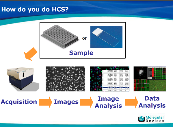
The MetaXpress XLS system (Molecular Devices, San Jose, CA, USA) is a high content analysis/screening platform which enables automatically multi-well plate/slide fluorescence imaging. It is composed of an imaging system with high sensitive, large field of viewing scientific CMOS camera for imaging acquisition in an automatic fashion with necessary computer hardware and software for image data management, data processing, analysis and bio-informatics for data mining and explorations. It is also equipped with associated hardware such as automated plate loader, liquid handling unit for increasing throughput.
|
System Aapplications:Although the system is designed as a screening platform using cellular response as a reporter to screen large number of chemical libraries, it is a high throughput fluorescence imaging platform which allows carrying out experiments in multi-well plate formats. It could be used for any fluorescence microscopy assays. The facility carries out routinely assays such as “transwell assay”, “wound healing assay”, “γH2AX foci counting assay” “Nanoparticle incorporation assay”, “live-dead assay”, etc. For a detailed application modules, please visit the MD web site: http://www.moleculardevices.com/applications;and check in the middle of the page: applications for a list of predefined modules.
The system is very flexible which allows creating customized modules for whatever fluorescence quantification applications you might need. The system is also equipped with phase optics and live cell imaging capability which allows performing fluorescence quantification with timelapse imaging. We have carried out live cell imaging for several days with 96 well plates. This allows increased throughput imaging which is difficult to carry out on a conventional fluorescence microscope. |
 |
Advantages of the MetaXpress system: The system has mainly 2 advantages over a conventional fluorescence microscope:high throughput and reliable fluorescence quantification. The system allows capturing of a large number of fluorescence images in an automatic fashion for quantification. The solid state light source of the system is highly stable which often a problem for a conventional fluorescence microscope where the light source intensity changes significantly over a short period of time.
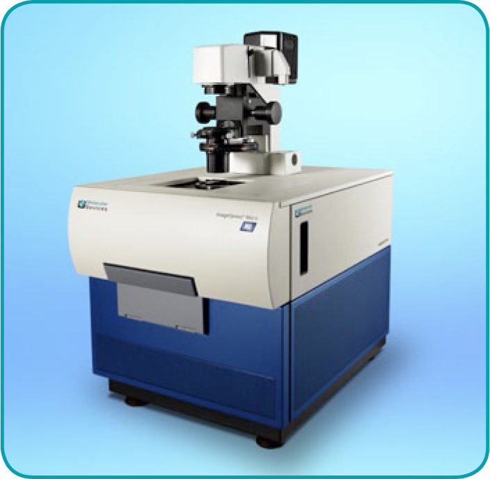
System description: The system is composed of a large field of view camera (2180X2180 at 6.5um per pixel), Nikon lenses, a solid state light source, a fast focusing motor, a high precision (100nm repeatability) stage and all necessary accessories (Temperature, CO2) for high speed live cell imaging. For a detailed description of the system, please visit MD website: http://www.moleculardevices.com/systems/high-content-imaging/imagexpress-micro-xls-widefield-high-content-analysis-system |
HCA/S System info:
Model: Molecular Devices MetaXpress XLS with plateloader
Objectives:
4X 0.2NA Plan-Apo
10X 0.3NA Plan-Apo
20X 0.75NA Plan-Apo
20X, 0.45NA S Plan Fluor ELWD
40X, 0.60NA S Pplan Fluor ELWD
40XPh2 0.60NA S Plan Fluor ELWD ADM
60X0.85NA Plan Fluor
Filter sets:
DAPI; FITC/ALEXA488; RHODAMINE/TRITC; CY3; TEXAS RED; CY5
Detectors
sCMOS camera 2180x2180, 6.5um pixel
DATA management and solution:
MD store, MD power core with 48 processors, AcuityXpress for bioinformatics
|
 |
|
|