|
|
|
| Equipment | LSM710 | Multiphoton STED | Spinning Disc Confocal | DIMs | PALM | FCS | FLIM | TEM | Micro-Injection | HCS | Workstataions and Tools |
Digital Imaging Microscopes (DIM)
DIMs offer alternative ways of imaging biological samples with advantages of low photo-bleaching, high-photon efficiency and relatively low cost. In combination with deconvolution, high resolution imaging could be performed on these systems.
|
 |
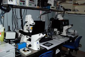
There are 6 DIM setups in the Facility. Each differs from the others by the software and hardware configuration for its main purposes:
- DIM station 1-4 contains Metamorph (Universal Imaging Corp , Molecular Devices), a sensitive, cooled CCD camera (Sensicam from Optikon , Cascade II EMCCD or CoolSnapHQ (Photometrics)) and a Zeiss microscope (AxioImagerZ, Axioplan IIM or Axiovert 200M). All of them are equipped with DIC (differential Interference Contrast) optics and fluorescence optics. The setups are mainly set for low-level fluorescent video microscopy. All these digital microscopes can perform multiple wavelength detection, 3-dimensional image acquisition and 3-D time lapse experiments. One setup is equipment with necessary filter wheels for Ca/pH ratiometric imaging and for FRET analysis with CFP and YFP dye pairs. It is also equipped with a live cell environment control that long term (days) timelapse can be performed on.
- DIM station 5 is composed of an Axioscope 2 from Zeiss for routine fluorescent examination and a high resolution digital color camera (Axiocam HR, Zeiss) for color digital photography.
- DIM station 6 is composed of a Zeiss Axiovert 100M, cooled CCD camera (Sensicam HE) and Metamorph software. The main difference between this setup and others is that this one is equipped with an Eppendorff microinjection system which allows injecting small amount of molecules in adherent cells. The system is, also available for routine fluorescence imaging.
- DIM station 7 contains an inverted fluorescence microscope (Olympus IX70) and a Zeiss Axiocam for routine sample examination as well as documentation. It could also perform time-lapse imaging with bright field (with phase contrast optics) if needed.
|
System info:
All these scopes (except 7) has interchangeable optics and are based on either upright or inverted configurations. Please contact staff for your particular application needs.
|
 |
|
|