Fluorescence Correlative Spectroscopy Module (Zeiss Confocor 2/3)
FCS is a spectroscopic technique for the study of molecular interactions in solution. FCS monitors the random motion of fluorescently labelled molecules inside a defined volume element irradiated by a focused laser beam. These fluctuations provide information on the rate of diffusion (diffusion time) of a particle and this, in turn, is directly dependent on the particle's mass. As a consequence, any increase in the mass of biomolecules, e.g. as a result of an interaction with a second molecule, is readily detected as an increase in the particle's diffusion time.
|
 |
Because FCS measurement is made through diffraction limited volume, FCS can be used to study molecule-molecule interactions in living cell. The system can be used to study:
- Receptor-ligand interactions
- Transcription factor-DNA interactions
- Lipid-protein interactions, etc.
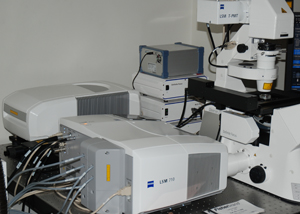
The FCS system at the Facility is integrated part of the Multi-photon and the LSM710/Zeiss LSM NLO systems with all the laser lines for common fluorophores and 2 detectors which enable cross correlation analysis.
More information about the system can be found on the following web site: http://www.zeiss.com/de/micro/home_e.nsf/Contents-FrameDHTML/4ACDCCD6CFEBFAEFC1256CB7004C8822
|
 |